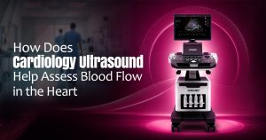
Cardiology ultrasound, commonly known as an echocardiogram, is generally considered an important test for assessing how well the heart is functioning. It uses safe sound waves to create live images of the heart as it beats. These images help doctors examine the heart chambers, valves, and pumping strength. Cardiovascular ultrasound also shows how blood flows through the heart and whether it is moving in the right direction. By accurately assessing blood flow, this test helps detect heart problems at an early stage.
What Is Cardiology Ultrasound?
Cardiology ultrasound is a non-invasive imaging test used to examine the structure and function of the heart. It is also sometimes referred to as an echosound for heart because it uses sound waves to create clear images of the beating heart. During the test, a small handheld device called a transducer is placed on the chest. This device sends sound waves into the body, and the returning echoes are converted into images on a screen. A human heart ultrasound helps doctors check the heart chambers, valves, and overall pumping function. In many cases, doctors combine regular ultrasound imaging with Doppler ultrasound, which helps measure the speed and direction of blood flow through the heart.
How Does It Assess Blood Flow?
The heart has four chambers and four valves that work together to move blood in the correct direction. An ultrasound of the heart is called an echocardiogram, and it helps doctors observe blood movement through the heart in real time. Doctors may recommend an ultrasound for heart problems to check whether blood is flowing normally, moving too slowly, moving too fast, or leaking backward.
- The direction of blood flow
- The speed of blood movement
- Blood flows across the heart valves
- Pressure changes inside the heart
- Abnormal flow patterns caused by defects or valve disease
Role in Checking Heart Valves
Heart valves open and close to keep blood flowing in one direction through the heart. Cardiac and vascular ultrasound helps doctors examine valve function and detect problems that may affect normal blood movement. Through cardiac ultrasound echocardiography, doctors can assess the four main valves and understand whether treatment or regular monitoring is needed.
- Valve stenosis: When a valve becomes narrowed and limits blood flow.
- Valve regurgitation: When a valve does not close properly, allowing blood to leak backward.
- Valve stiffness: When a valve does not open or close smoothly.
- Valve damage: When infection, age, or disease affects normal valve function.
Detecting Abnormal Blood Flow Patterns
Abnormal blood flow can be a sign of several heart conditions. An ultrasound of the heart, called an echocardiogram that may help identify problems such as congenital heart defects, valve disease, heart failure, cardiomyopathy, and pressure changes in the heart or lungs. This echocardiogram test allows doctors to study blood movement and detect changes that may affect normal heart function. For example, if blood is moving faster than expected through a valve, it may suggest narrowing. If blood is flowing backward, it may indicate valve leakage. If the heart is not pumping strongly, the ultrasound can show reduced movement of the heart muscle and changes in blood flow.
Why Doctors Recommend This Test
Doctors may suggest a cardiology ultrasound if a patient has symptoms such as chest pain, shortness of breath, swelling in the legs, dizziness, irregular heartbeat, or a heart murmur. Since an ultrasound of the heart is called an echocardiogram, doctors often use it to monitor existing heart conditions or check how well a treatment is working. In some cases, doctors may recommend an ultrasound of the heart valves to identify valve narrowing, leakage, or abnormal blood flow. It helps doctors make better decisions about medications, lifestyle changes, further testing, or procedures.
Benefits of Cardiology Ultrasound
- Non-invasive and generally painless testing
- No radiation exposure
- Real-time images of the beating heart
- Detailed assessment of blood flow
- Helpful in diagnosing valve and pumping problems
- Useful for monitoring heart conditions over time
A Healthier Heart Starts with Better Understanding
Cardiology ultrasounds always have an important role in assessing blood flow and overall heart function. By using echocardiography and Doppler technology, doctors can see how blood moves through the heart chambers and valves. A sonogram of the heart valves can help detect abnormal flow patterns, congenital defects, and other cardiovascular conditions easily. For people experiencing breathlessness, chest discomfort, palpitations, or unexplained fatigue, this test can provide important answers about heart health. With early diagnosis and proper medical guidance, many heart concerns can be managed more effectively. Overall, cardiology ultrasound is a safe test that supports better heart care and helps patients take confident steps toward a healthier future.