
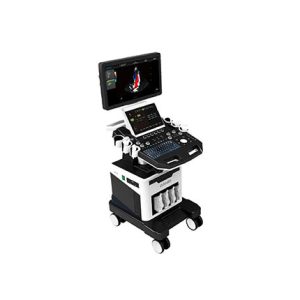
Light As Swallow
Accurate And Comprehensive
1.Dual probe socket, portable color Doppler ultrasound system
2.Using a new computing engine and image processing technology
3.Has a sensitive blood flow detection capability and a wide range of probe adaptation capabilities
4.Meet the abdomen, obstetrics and gynecology, heart, small organs, superficial blood vessels, musculoskeletal, four-dimensional, etc.
Except routine examination needs, can also meet the clinical needs of other specialized clinics.
Trapezoidal imaging
Refers to converting the line data of a linear array probe into a trapezoidal image through coordinate transformation and interpolation, which is an extended imaging.
Freehand elastography mode
Freehand elastography can help doctors distinguish between soft/hard lesions and surrounding tissues.
Wide-field imaging
Also known as ultra-wide-field imaging. Compared with ordinary ultrasound imaging, wide-field imaging provides a new perspective for clinical diagnosis, and has a very important clinical significance for observing large lesions and the relationship between the lesions and surrounding tissues. Diagnostic significance.
Contrast imaging
The use of significantly different echo charac teristics from human soft tissue, or acoustic characteristic impedance (i.e., specific acous tic impedance).
Anatomical M-mode
Has only one m-sampling line, which has limitations for moving examination tissues, especially for difficult patients. The anatomical M-mode makes up for the lack of traditional M-modes for the examination of patients with difficult imaging, and it provides multiple M-sampling lines. To enable you to perform more effective motion analysis on M-mode images at different angles and positions.
Puncture enhancement
Automatic detection of the needle body, automatic deflection of the sound beam, and smart puncture enhancement technology make the puncture display in the human body more intuitive.
Color M mode
Abbreviated as MC mode, used for cardiac examination applications. Color blood flow uses speed and variance color maps to make colors superimposed on M-mode images. Color blood flow covers B-mode images and M-mode timeline.
D mode
Also known as PW mode. PW Doppler allows you to selectively check blood flow data from a small area (i.e. the sampling volume). PW Doppler displays blood flow information through a constantly moving spectrum, visually Describes the functional relationship between blood flow velocity and time
Automatic IMT measurement
The thickness of the intima of the blood vessel is an important indicator
for predicting the risk of cardiovascular disease. The automatic measurement technique of the intima can automatically measure the thickness
of the intima in the near and far fields of the blood vessel and automatically optimize the measurement angle.
15-Inch full-view medical HD display on the main screen
USB 3.0 interface clinical picture, video, report storage, export
Real-time wide-field imaging (WFOV)
Trapezoidal imaging, continuous Doppler imaging
Free anatomy 3M imaging
IMT automatic measurement of blood vessel intima
Free arm 3D imaging, real time 4D imaging
Spatial composite imaging speckle noise removal technology
Tissue Doppler imaging (TDI)
2 reviews for CARDIOLOGY ULTRASOUND MACHINE – PORTABLE
There are no reviews yet.