
High quality clinical performance: space composite imaging, one-button image solution to greatly improve work efficiency, the use of real-time volume probe, and mainstream imaging technology, further shorten the diagnostic distance.
Stable and efficient: deeply optimized Windows operating platform and integrated high-performance and low-power industrial control solutions.
User-friendly operation design: touch screen fast navigation, reduce button contact and press, simplify operation steps, reduce user fatigue to a greater extent, and improve the work efficiency of doctors.
High-cost performance: all software functions are opened once, and additional consumption is refused; Full service tracking.
M – measuring cardiac function –
The system provides a variety of automatic measurement tools, so that the operation is simple, fast and accurate.
Tissue Doppler – cardiac muscle movement –
The adaptive signal processing technology is adopted to analyze the echo signal in a specific area through the unique intelligent data perception method, so as to improve the image resolution and uniformity and easily obtain the high-definition heart image.
Specifications –
Operating system: Windows 7 operating system
Application range :
Abdomen, obstetrics, gynecology, heart, urinary system, small organs, superficial, blood vessels, pediatrics, newborns, muscles, etc.
Probes:
Abdomen, obstetrics, gynecology, heart, urinary system, small organs, superficial, blood vessels, pediatrics, newborns, muscles, etc.
Measurements and reports:
Support advanced measurement software packages, report software packages, case management software packages, etc. for abdomen, obstetrics, gynecology, heart, urinary system, small organs, superficial, vascular, pediatric, etc.
IMT mesurements
Automatic spectrum envelope measurement
All-digital transmit and receive beamformer
Color Doppler Imaging (C)
Pulse Doppler Imaging (PW)
Contrast imaging (CTI)
Continuous Wave Doppler Imaging (CW)
B/C/D triple synchronous real-time imaging
Power Doppler Imaging (PDI)
Directional Power Doppler (DPDI)
M mode imaging
Anatomical M imaging
Blood flow M (MC) imaging
Freehand elastography
Tissue Doppler Imaging (TDI )
Strain rate imaging
Harmonic Imaging Imaging (THI)
Harmonic fusion imaging (FHI)
Adaptive Speckle Noise Suppression (SRI)
Panoramic Imaging
Deflection imaging
Trapezoidal imaging
Adaptive sound speed optimization
Freehand 3D imaging
Real-time four-dimensional imaging (3D/4D)
DICOM3.0
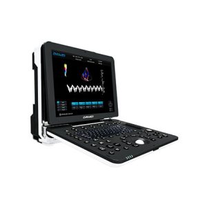
Monitor: 21.5 inches, high-resolution ultrasound dedicated LED
13.3 inch touch screen
Integrated clipboard: Saved images are displayed at the bottom of the screen and can be directly transferred or deleted
System with field upgrade function
Presets: pre-select optimized inspection conditions for different inspection organs, reduce adjustment during operation, and commonly used external adjustments and combination adjustments
Probe interface ≥ 4
English operation interface
Detection depth: ≥360mm
Extended imaging
Reviews
There are no reviews yet.